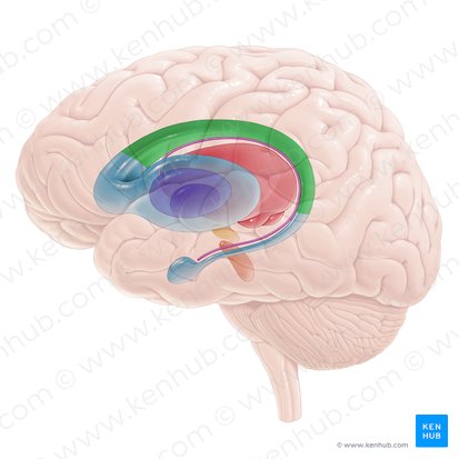
Caudate nucleus
At the time the article was last revised Yoshi Yu had no financial relationships to ineligible caudate nucleus to disclose. Caudate nuclei are paired nuclei which along with the globus pallidus and putamen are referred to as the corpus striatumand collectively make up the basal ganglia. The caudate nuclei have both motor and behavioral functions, in particular maintaining body and limb posture, as well as controlling approach-attachment behaviors, respectively 3, caudate nucleus. The caudate caudate nucleus is located lateral to the lateral ventricles, with the head lateral to the frontal horn, and body lateral to the body of the lateral ventricle.
Federal government websites often end in. Before sharing sensitive information, make sure you're on a federal government site. The site is secure. NCBI Bookshelf. Margaret E.
Caudate nucleus
Deep within each half of the brain lies the caudate nucleus. The caudate nucleus is a pair of brain structures that make up part of the basal ganglia. It helps control high-level functioning, including:. The basal ganglia are neuron cell bodies found deep within the brain involved with movement, behavior, and emotions. This brain circuit receives information from the cerebral cortex, which is a layer of grey matter in the outer brain linked to higher cognitive functions such as information processing and learning. The basal ganglia sends information mainly to the thalamus , which sends information back to the cerebral cortex. The nuclei feature a wide head that tapers into a body and a thin tail. The caudate nucleus helps process visual information and control movement. The structure plays a vital role in how the brain learns, specifically the storing and processing of memories. As a feedback processor, it uses information from past experiences to influence future actions and decisions. This is important to the development and use of language. Experts think that communication skills are controlled mostly by the caudate nucleus and the thalamus. Another brain structure called the substantia nigra releases dopamine that projects to the caudate nucleus.
Warrant EJ ed.
It plays a critical role in various higher neurological functions. Each caudate nucleus is composed of a large anterior head, a body, and a thin tail that wraps anteriorly such that the caudate nucleus head and tail can be visible in the same coronal cut. When combined with the putamen, the pair is referred to as the striatum and is often considered jointly in function. The striatum is the major input source for the basal ganglia, which also includes the globus pallidus, subthalamic nucleus, and substantia nigra. These deep brain structures together largely control voluntary skeletal movement. The caudate nucleus functions not only in planning the execution of movement, but also in learning, memory, reward, motivation, emotion, and romantic interaction. Input to the caudate nucleus travels from the cortex, mostly the ipsilateral frontal lobe.
The caudate nuclei there are two—one on each side of the brain can be found below the cerebral cortex , situated next to the lateral ventricles. Like the lateral ventricles, the caudate is a C-shaped structure with a thick anterior portion called the head , which becomes narrower as it extends towards the back of the brain. The middle portion of the caudate is known as the body , and this tapers off into the tail of the caudate. The caudate nucleus is considered part of the basal ganglia. The basal ganglia are a group of subcortical nuclei that are involved in a variety of cognitive and emotional functions, but are best known for their role in movement.
Caudate nucleus
At the time the article was last revised Yoshi Yu had no financial relationships to ineligible companies to disclose. Caudate nuclei are paired nuclei which along with the globus pallidus and putamen are referred to as the corpus striatum , and collectively make up the basal ganglia. The caudate nuclei have both motor and behavioral functions, in particular maintaining body and limb posture, as well as controlling approach-attachment behaviors, respectively 3. The caudate nucleus is located lateral to the lateral ventricles, with the head lateral to the frontal horn, and body lateral to the body of the lateral ventricle.
Floating corner shelves white
Dingman explores some of the most fascinating and mysterious expressions of human behavior in a style that is case study, dramatic novel, and introductory textbook all rolled into one. Nat Neurosci. The caudate nucleus is considered part of the basal ganglia. While the correlation does not indicate causation, the finding may have implications for early diagnosis. Recent Edits. Karger AG. Differential abnormalities of the head and body of the caudate nucleus in attention deficit-hyperactivity disorder. Article created:. Know Your Brain: Basal Ganglia. StatPearls [Internet]. Prog Neurobiol. Why do we have a caudate nucleus?. Normal, nonpathological differences in the caudate nucleus occur along the lines of age and genetic and environmental exposures. Lesions in the dorsolateral caudate nucleus predict post stroke depression — a voxel-based lesion-symptom mapping study. The body of fornix joins the hippocampus and mammillary bodies, structures in the base of the brain that are involved in memory formation and recall….
The caudate nucleus is one of the structures that make up the corpus striatum , which is a component of the basal ganglia in the human brain. The caudate is also one of the brain structures which compose the reward system and functions as part of the cortico — basal ganglia — thalamic loop.
The caudate nucleus integrates visual input and inhibits the substantia nigra, disinhibiting the superior colliculus to enable the coordination of eye movement, and is important in voluntary saccadic eye movement. URL of Article. The only organ system lacking a lymphatic system is the central nervous system, which was previously believed to lack lymphatic drainage completely. Close Please Note: You can also scroll through stacks with your mouse wheel or the keyboard arrow keys. The patient maintained language comprehension in her three languages, but when asked to produce language, she involuntarily switched between the three languages. Sexual dimorphism of brain developmental trajectories during childhood and adolescence. Turn recording back on. Last revised:. In Parkinson disease, the dopaminergic innervation of the caudate and putamen is severely compromised by the death of dopamine neurons in the substantia nigra pars compacta Chapter Cortical spreading depression modulates the caudate nucleus activity. Wikimedia Commons.


At me a similar situation. I invite to discussion.
Certainly. All above told the truth.