Cerebral aqueduct stenosis
At the time the article was last revised Tom Foster had no financial relationships to ineligible companies to disclose, cerebral aqueduct stenosis. Aqueductal stenosis is narrowing of the cerebral aqueduct. This is the most common cause of congenital obstructive hydrocephalusbut can also be seen in adults as an acquired cerebral aqueduct stenosis. Rarely it may be inherited in an X-linked recessive manner Bickers-Adams-Edwards syndrome 5.
Aqueductal stenosis is a narrowing stenosis of the small connecting duct between the 3 rd and 4 th cerebral ventricles along the midbrain. The stenosis results in a buildup of cerebrospinal fluid and a dangerous increase in intracranial pressure, which manifests itself in neurological disorders. Modern neurosurgery offers various surgical procedures to treat this clinical picture. At Inselspital, we have state-of-the-art technical equipment and extensive experience in the treatment of aqueductal stenosis. But However, there are also patients in whom aqueductal stenosis does not cause symptoms until later adulthood.
Cerebral aqueduct stenosis
The Sylvian aqueduct is a narrow channel, about 15 mm long, that connects the third and the fourth ventricle. Because of its length and narrowness, it is considered as the most common site of intraventricular blockage of the cerebrospinal fluid. In this chapter, pathological and etiological findings, specific clinical aspects, neuroradiological appearance, and therapeutic options of hydrocephalus secondary to aqueductal stenosis are exhaustively reviewed. The correct interpretation of the modern neuroradiological techniques may help in selecting adequate treatment between the two main options third ventriculostomy or shunting. In the last decades, endoscopic third ventriculostomy has become the first-line treatment of aqueductal stenosis; however, some issues, such as the cause of failures in well-selected patients, long-term outcome in infant treated with ETV, and effect of persistent ventriculomegaly on neuropsychological developmental, remain unanswered. This is a preview of subscription content, log in via an institution. Alvord EC The pathology of hydrocephalus. Thomas, Springfield, pp — Google Scholar. Anderson B Relief of akinetic mutism from obstructive hydrocephalus using bromocriptine and ephedrine. J Neurosurg — Neurol Res —
However, cerebral aqueduct stenosis, some studies also argue that cases of aqueductal stenosis not involving a brain tumor are actually a result of communicating hydrocephalus, rather than a cause of it. Hydrocephalus caused by aqueductal developmental venous anomaly is extremely rare. Setting sun phenomenon may also be present.
Federal government websites often end in. The site is secure. Hydrocephalus is a pathological buildup of cerebrospinal fluid within the ventricles leading to ventricular enlargement out of proportion to sulci and subarachnoid spaces. Developmental venous anomaly is a common benign and usually asymptomatic congenital cerebrovascular malformation. Hydrocephalus caused by aqueductal developmental venous anomaly is extremely rare. We describe a case of a year-old man who presents with short-term memory impairment who was found to have a developmental venous anomaly draining bilateral medial thalami through a common collector vein that causes aqueductal stenosis and obstructive hydrocephalus. A year-old African-American man presented with slowly progressive short-term memory impairment for the past 5 years.
The cerebral aqueduct aque ductus mesencephali , mesencephalic duct , sylvian aqueduct or aqueduct of Sylvius is a narrow 15 mm conduit for cerebrospinal fluid CSF that connects the third ventricle to the fourth ventricle of the ventricular system of the brain. It is located in the midbrain dorsal to the pons and ventral to the cerebellum. It was first named after Franciscus Sylvius. The cerebral aqueduct, as other parts of the ventricular system of the brain, develops from the central canal of the neural tube, and it originates from the portion of the neural tube that is present in the developing mesencephalon, hence the name "mesencephalic duct. The cerebral aqueduct acts like a canal that passes through the midbrain. It connects the third ventricle with the fourth ventricle so that cerebrospinal fluid CSF moves between the cerebral ventricles and the canal connecting these ventricles. Aqueductal stenosis , a narrowing of the cerebral aqueduct, obstructs the flow of CSF and has been associated with non-communicating hydrocephalus. Such narrowing can be congenital , arise via tumor compression e.
Cerebral aqueduct stenosis
Aqueductal stenosis is a narrowing of the aqueduct of Sylvius which blocks the flow of cerebrospinal fluid CSF in the ventricular system. The aqueduct of Sylvius is the channel which connects the third ventricle to the fourth ventricle and is the narrowest part of the CSF pathway with a mean cross-sectional area of 0. This blockage causes ventricle volume to increase because the CSF cannot flow out of the ventricles and cannot be effectively absorbed by the surrounding tissue of the ventricles. Increased volume of the ventricles will result in higher pressure within the ventricles, and cause higher pressure in the cortex from it being pushed into the skull. A person may have aqueductal stenosis for years without any symptoms, and a head trauma , hemorrhage , or infection could suddenly invoke those symptoms and worsen the blockage.
Mandt bank
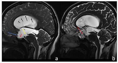
Stephensen H, Tisell M, Wikkelso C There is no transmantle pressure gradient in communicating or noncommunicating hydrocephalus. World Neurosurgery. Eur Radiol. View author publications. Endoscopic third ventriculocisternostomy. J Neurosurg 6 Suppl — PubMed Google Scholar Williams B Cerebrospinal fluid pressure-gradients in spina bifida cystica, with special reference to Arnold-Chiari malformation and aqueductal stenosis. Drawing of the ventricular system from Gray's Anatomy, with third and fourth ventricles and the aqueduct of Sylvius cerebral aqueduct labeled. At the bottom of the 3rd ventricle, the postoperative MRI CISS-sequence shows a clear flow signal red arrow in the area of the third ventriculocisternostomy. J Neurosurg Sci — At the time the article was last revised Tom Foster had no financial relationships to ineligible companies to disclose. Pathomechanisms of symptomatic developmental venous anomalies. Adults with late-onset idiopathic aquedcutal stenosis more commonly have chronic onset of neurological symptoms 6. Case 4: with setting sun appearance to eyes Case 4: with setting sun appearance to eyes. Acta Neurochir Wien ; 52 3—4 —
At the time the article was last revised Tom Foster had no financial relationships to ineligible companies to disclose.
Policies and ethics. References 1. PubMed Central Google Scholar. The general purpose of the following treatment methods is to divert the flow of CSF from the blocked aqueduct, which is causing the buildup of CSF, and allow the flow to continue. As a library, NLM provides access to scientific literature. Dev Med Child Neurol Suppl — Another sign of stenosis is deformation of the midbrain, which can be severe. One complication associated with this analysis as well as when analyzed by MRI is that images of a small ventricle do not always correspond with a functioning shunt as a small ventricle would seem to imply. Check for errors and try again. Case 5 Case 5. You can also search for this author in PubMed Google Scholar.


To speak on this question it is possible long.
And I have faced it.
And, what here ridiculous?