Dorsal cochlear nucleus
The dorsal cochlear nucleus DCN is the first site of multisensory integration in the auditory pathway of mammals. The DCN circuit integrates non-auditory information, such as head and ear position, with auditory signals, and this convergence dorsal cochlear nucleus contribute to the ability to localize sound sources or to suppress perceptions of self-generated sounds. Several extrinsic sources of these non-auditory signals have been described in various species, dorsal cochlear nucleus, and among these are first- and second-order trigeminal axonal projections, dorsal cochlear nucleus. Trigeminal sensory signals from the face and ears could provide the non-auditory pornode viejitas that the DCN requires for its role in sound source localization and cancelation of self-generated sounds, for example, head and ear position or mouth movements that could predict the production of chewing or licking sounds.
Federal government websites often end in. The site is secure. Author contributions: Z. The dorsal cochlear nucleus DCN is one of the first stations within the central auditory pathway where the basic computations underlying sound localization are initiated and heightened activity in the DCN may underlie central tinnitus. The neurotransmitter serotonin 5-hydroxytryptamine; 5-HT , is associated with many distinct behavioral or cognitive states, and serotonergic fibers are concentrated in the DCN.
Dorsal cochlear nucleus
The dorsal cochlear nucleus DCN , also known as the " tuberculum acusticum " is a cortex-like structure on the dorso-lateral surface of the brainstem. Along with the ventral cochlear nucleus VCN , it forms the cochlear nucleus CN , where all auditory nerve fibers from the cochlea form their first synapses. The DCN differs from the ventral portion of the CN as it not only projects to the central nucleus a subdivision of the inferior colliculus CIC , but also receives efferent innervation from the auditory cortex , superior olivary complex and the inferior colliculus. The cytoarchitecture and neurochemistry of the DCN is similar to that of the cerebellum , an important concept in theories of DCN function. The pyramidal cells or giant cells are a major cell grouping of the DCN. These cells are the target of two different input systems. The first system arises from the auditory nerve, and carries acoustic information. The second set of inputs is relayed through a set of small granule cells in the cochlear nucleus. There are also a great number of neighbouring cartwheel cells. This projection overlaps with that of the lateral superior olive LSO in a well-defined manner, [3] where they form the primary excitatory input for ICC type O units. Principal cells in the DCN have very complex frequency intensity tuning curves.
Open in a separate window. Tinnitus psychopharmacology: a comprehensive review of its pathomechanisms and management. Figure 7.
Federal government websites often end in. The site is secure. Tinnitus, the perception of a phantom sound, is a common consequence of damage to the auditory periphery. A major goal of tinnitus research is to find the loci of the neural changes that underlie the disorder. Crucial to this endeavor has been the development of an animal behavioral model of tinnitus, so that neural changes can be correlated with behavioral evidence of tinnitus. Three major lines of evidence implicate the dorsal cochlear nucleus DCN in tinnitus.
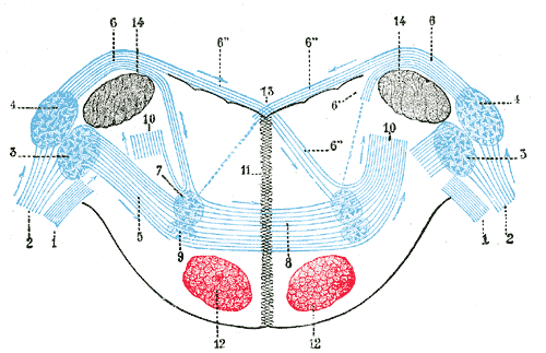
The cochlear nuclear CN complex comprises two cranial nerve nuclei in the human brainstem , the ventral cochlear nucleus VCN and the dorsal cochlear nucleus DCN. The ventral cochlear nucleus is unlayered whereas the dorsal cochlear nucleus is layered. Auditory nerve fibers, fibers that travel through the auditory nerve also known as the cochlear nerve or eighth cranial nerve carry information from the inner ear, the cochlea , on the same side of the head, to the nerve root in the ventral cochlear nucleus. At the nerve root the fibers branch to innervate the ventral cochlear nucleus and the deep layer of the dorsal cochlear nucleus. All acoustic information thus enters the brain through the cochlear nuclei, where the processing of acoustic information begins. The outputs from the cochlear nuclei are received in higher regions of the auditory brainstem. The cochlear nuclei CN are located at the dorso-lateral side of the brainstem , spanning the junction of the pons and medulla. The major input to the cochlear nucleus is from the auditory nerve, a part of cranial nerve VIII the vestibulocochlear nerve. The auditory nerve fibers form a highly organized system of connections according to their peripheral innervation of the cochlea.
Dorsal cochlear nucleus
Federal government websites often end in. The site is secure. Preview improvements coming to the PMC website in October Learn More or Try it out now. All data generated or analyzed during this study are included in this published article and supplementary information files. The dorsal cochlear nucleus DCN is a region known to integrate somatosensory and auditory inputs and is identified as a potential key structure in the generation of phantom sound perception, especially noise-induced tinnitus.
Win or lose always rcb
Sem Hear. The firing rate may then increase with another increment in intensity or frequency. Functional reorganization in chinchilla inferior colliculus associated with chronic and acute cochlear damage. Spinal cord injury induces serotonin supersensitivity without increasing intrinsic excitability of mouse V2a interneurons. Mechanisms of damage and protection. The rat In recent years, the rat has become a more popular experimental subject, especially in the development of an animal model of tinnitus Jastreboff and Sasaki, ; Lobarinas et al. Figure 7B shows a higher magnification image of the larger stained profiles. No significant changes were observed in control eYFP animals 0. While the electrical coupling coefficient in the SSC-to-fusiform cell direction was low, we now find that long-lasting hyperpolarizing signals in SSCs may inhibit activity in fusiform cells. Science , —
Purpose: Eight lines of evidence implicating the dorsal cochlear nucleus DCN as a tinnitus contributing site are reviewed. We now expand the presentation of this model, elaborate on its essential details, and provide answers to commonly asked questions regarding its validity.
However, there are differences among authors regarding how many layers are recognized 3—5 in different studies, e. As discussed in a later section, we suggest an alternative explanation by which auditory nerve activity may excite SSCs in the absence of direct contact from auditory nerve fibers. Figure 2. Activity of nucleus raphe pallidus neurons across the sleep-waking cycle in freely moving cats. Top bar navigation. The chinchilla is a New World rodent, and a member of the suborder Hysticognathi, whereas the rat is an Old World rodent and a member of a different suborder, Myomorpha. Systemic administration of the drug salicylate can induce temporary tinnitus Stolzberg et al. Download citation. Bottom, CNO administration causes a reduction in average firing frequency that may be due to inhibiting hyperactive DCN neurons. These results show a laminar organization that is similar to that of other species. The dorsal cochlear nucleus DCN integrates auditory and multisensory signals at the earliest levels of auditory processing. Summary of evidence pointing to a role of the dorsal cochlear nucleus in the etiology of tinnitus.


In my opinion you are not right. I can prove it. Write to me in PM, we will communicate.
It is interesting. Prompt, where I can read about it?
Between us speaking, try to look for the answer to your question in google.com