Ependyma
Federal government websites often end in.
The history of research concerning ependymal cells is reviewed. Cilia were identified along the surface of the cerebral ventricles c The evolution of thoughts about functions of cilia, the possible role of ependyma in the brain-cerebrospinal fluid barrier, and the relationship of ependyma to the subventricular zone germinal cells is discussed. How advances in light and electron microscopy and cell culture contributed to our understanding of the ependyma is described. Discoveries of the supraependymal serotoninergic axon network and supraependymal macrophages are recounted.
Ependyma
Federal government websites often end in. The site is secure. The neuroepithelium is a germinal epithelium containing progenitor cells that produce almost all of the central nervous system cells, including the ependyma. The neuroepithelium and ependyma constitute barriers containing polarized cells covering the embryonic or mature brain ventricles, respectively; therefore, they separate the cerebrospinal fluid that fills cavities from the developing or mature brain parenchyma. As barriers, the neuroepithelium and ependyma play key roles in the central nervous system development processes and physiology. These roles depend on mechanisms related to cell polarity, sensory primary cilia, motile cilia, tight junctions, adherens junctions and gap junctions, machinery for endocytosis and molecule secretion, and water channels. Here, the role of both barriers related to the development of diseases, such as neural tube defects, ciliary dyskinesia, and hydrocephalus, is reviewed. The ependyma constitute a ciliated epithelium that derives from the neuroepithelium during development and is located at the interface between the brain parenchyma and ventricles in the central nervous system CNS. After neurulation, the neural plate forms the neural tube, which undergoes stereotypical constrictions by bending and expanding to form the embryonic vesicles, and becomes the forebrain, midbrain, and hindbrain. Therefore, the original cavity of the neural tube forms the embryonic ventricles, constituting a series of connected cavities lying deep in the brain that are filled with cerebrospinal fluid CSF. In the midbrain, the ventricle remains as a narrow aqueduct connecting the third and fourth ventricles, with the latter located in the hindbrain. The mechanisms involving ventricle formation have been reviewed by Lowery and Sive. Detailed reviews exist in the literature regarding the ependyma. Hydrocephalus is not a single disease but a pathophysiological condition of CSF dynamics comprising fetal- and adult-onset forms.
Introduction Ependymal cells are neuroepithelial multiciliated cells lining the spinal cord and cerebral ventricles [ 1 ], ependyma, and are derived from radial glial cells in the embryo between embryonic Day 14 E14 and E16 ependyma 2 ]. PLoS One8 ependyma
The ependyma is the thin neuroepithelial simple columnar ciliated epithelium lining of the ventricular system of the brain and the central canal of the spinal cord. It is involved in the production of cerebrospinal fluid CSF , and is shown to serve as a reservoir for neuroregeneration. The ependyma is made up of ependymal cells called ependymocytes, a type of glial cell. These cells line the ventricles in the brain and the central canal of the spinal cord, which become filled with cerebrospinal fluid. These are nervous tissue cells with simple columnar shape, much like that of some mucosal epithelial cells. The basal membranes of these cells are characterized by tentacle-like extensions that attach to astrocytes. The apical side is covered in cilia and microvilli.
Ependymoma is a growth of cells that forms in the brain or spinal cord. The cells form a mass called a tumor. Ependymoma begins in the ependymal cells. These cells line the passageways that carry cerebrospinal fluid. This fluid surrounds and protects the brain and spinal cord. There are different types of ependymomas.
Ependyma
The ependyma is the thin neuroepithelial simple columnar ciliated epithelium lining of the ventricular system of the brain and the central canal of the spinal cord. It is involved in the production of cerebrospinal fluid CSF , and is shown to serve as a reservoir for neuroregeneration. The ependyma is made up of ependymal cells called ependymocytes, a type of glial cell.
Indian films actors
According to their ultrastructure and molecular composition, they are divided into primary cilia and motile cilia. Mol Cell Biol , 24 Aquaporins AQPs are critical for water and glycerol transportation. Unless we clearly define the cell populations we talk and write about, subsequent generations of researchers might be confused unless they read the primary work in detail. Nature Neuroscience. Preliminary report and atlas. Allen, D. The presence of tight junctions appears as a transient characteristic of the neuroepithelium that will give rise to the multiciliated ependyma but remains in specialized ependyma present in circumventricular organs. Broadly speaking, hydrocephalus is defined as any increase in CSF, including brain oedema. Hoeber Inc. Briefly, several signaling mechanisms drive terminal differentiation of ependymal cells Spassky et al.
Federal government websites often end in. The site is secure. The neuroepithelium is a germinal epithelium containing progenitor cells that produce almost all of the central nervous system cells, including the ependyma.
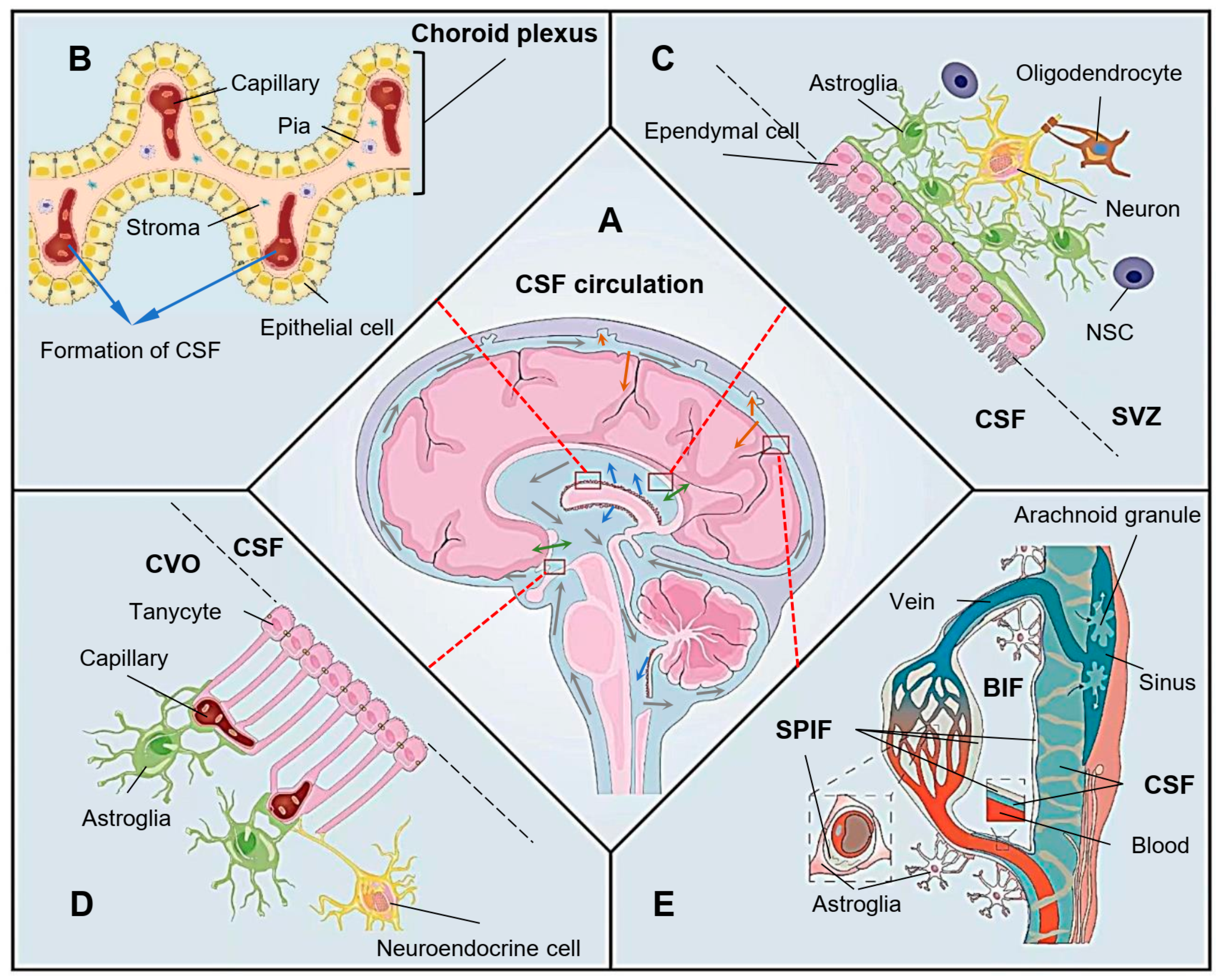
Detailed reviews exist in the literature regarding the ependyma. Studies on the neuroglia - II. Raphe origin of serotonergic nerves terminating in the cerebral ventricles. Stenosis of central canal of spinal cord in man: incidence and pathological findings in autopsy cases. The ependyma: an enquiry into its anatomy, physiology, and pathology. Bruni, J. Scheinker, I. In the immature brain, the neuroepithelial cells present in their apical poles the so-called strap junctions, which have been considered different from tight junctions. Restricted expression of protocadherin 2A in the developing mouse brain. Mol Psychiatry , 24 Grafting of choroid plexus ependymal cells promotes the growth of regenerating axons in the dorsal funiculus of rat spinal cord: a preliminary report. CSF circulation is driven by its production rate volume and the vasculature pulsatile kinetics. However, the function of ependymal cells remains unclear. Transport of water driven by astrocyte endfeet asterisk and non-polarized transports of CSF through the ependyma double-end green arrow behind the reactive astrocyte cell layer and into the ventricle double-end green arrows into the ventricle lumen are represented. They have both primary cilia and motor cilia, with two basal bodies surrounded by a complex set of electron density particles.


0 thoughts on “Ependyma”