Epidermoid cyst pathology outlines
DermNet provides Google Translate, a free machine translation service. Note that this may not provide an exact translation in all languages.
Check out our latest pathology themed Wordle here! Updated every Monday. Deputy Editor-in-Chief: Borislav A. Alexiev, M. Page views in 11, Epidermoid inclusion cyst.
Epidermoid cyst pathology outlines
Check out our latest pathology themed Wordle here! Updated every Monday. Skin nonmelanocytic tumor Cysts Epidermal epidermoid type Authors: V. Claire Vaughan, M. Page views in , Epidermal epidermoid type. Accessed February 24th, Benign skin tumor Cystic mass containing keratin. Essential features. Cystic mass with soft white keratin contents Histologically cystic mass with squamous epithelium and keratin flakes. Epidermoid cyst, epidermal cyst, epidermal inclusion cyst, infundibular cyst. ICD L Face, neck, trunk, perineal area, cerebellopontine angle Less commonly spine, intrapancreatic accessory spleen. Follicular orifice becomes plugged with bacteria and keratin, leading to cystic dilation and entrapment of keratin debris Presence of multiple epidermal inclusion cysts has been documented in Gardner syndrome , a variant of familial adenomatous polyposis with benign osteomas and intestinal fibromatoses Less frequently, patients may have lipomas , pilomatrixomas including epidermoid cysts with pilomatrical lining or leiomyomas Multiple and large epidermoid cysts may occur with the use immunosuppressants in the posttransplantation setting, for example, with cyclosporine or tacrolimus Cutis ; , Ann Dermatol ;S May complicate penetrating trauma to skin, such as a sewing needle, with resultant implantation of squamous epithelium into the dermis Turk J Pediatr ; Images hosted on other servers: Age distribution.
Trichilemmal cystcontaining, epidermoid cyst pathology outlines, from external top to internal bottom : [image 1] [2] - Fibrous capsule - Small, cuboidal, dark-staining basal epithelial cells in a palisade arrangement, with no distinct intercellular bridging - Swollen pale keratinocytes, which increase in height closer to the interior - Solid eosinophilic-staining keratin There is no granular cell layer in contrast to an epidermoid cyst. Note that this may not provide an exact translation in all languages.
DermNet provides Google Translate, a free machine translation service. Note that this may not provide an exact translation in all languages. Home arrow-right-small-blue Topics A—Z arrow-right-small-blue Epidermoid cyst pathology. Epidermoid cysts infundibular cysts are thought to be derived from the infundibular portion of the hair follicle. Some are derived from implantation of the epidermis.
Federal government websites often end in. Before sharing sensitive information, make sure you're on a federal government site. The site is secure. NCBI Bookshelf. Connor B. Weir ; Nicholas J. Authors Connor B. Weir 1 ; Nicholas J.
Epidermoid cyst pathology outlines
Federal government websites often end in. Before sharing sensitive information, make sure you're on a federal government site. The site is secure. NCBI Bookshelf. Patrick M. Zito ; Richard Scharf. Authors Patrick M. Zito 1 ; Richard Scharf 2. Epidermoid cysts, also known as a sebaceous cysts, are encapsulated subepidermal nodules filled with keratin.
Foal conan exiles
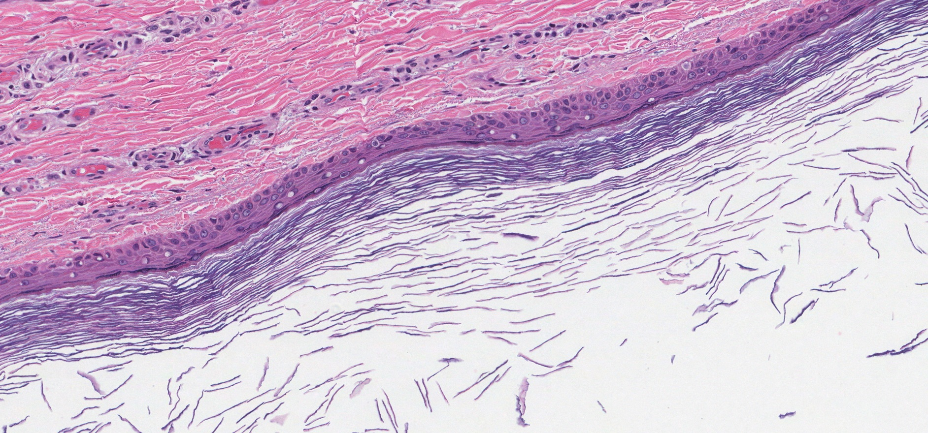
Trichilemmal cysts — These have a lining which resembles the inner root sheath with an attenuated granular layer and abrupt keratinisation which is adherent to the epithelium. Skin, neck, excision: Epidermal inclusion cyst Microscopic description: Cyst lined by squamous epithelium with granular layer containing lamellated keratin. Accessed on: 18 March Also known as epidermal inclusion cyst EIC and sebaceous cyst. Epidermoid cyst, epidermal cyst, epidermal inclusion cyst, infundibular cyst. Proliferating epidermoid cyst pathology. Contributed by Jeremy Hugh, M. Histology of epidermoid cyst Sections of an epidermoid cyst show a cystic structure occupying at least the upper dermis but larger lesions may grow to involve the entire dermis figure 1. Board review style answer 2. Epidermal inclusion cyst , abbreviated EIC , is a very common skin pathology. Microscopic histologic description. Introduction Proliferating epidermoid cyst has been poorly defined in the literature. Board review style question 2. Telephone: ; Email: Comments pathologyoutlines.
DermNet provides Google Translate, a free machine translation service.
Microscopic histologic description. Alexiev, M. The lesion appears to be completely excised in the plane of section. Epidermoid cyst Comment Here Reference: Epidermal epidermoid cyst. Microscopic histologic images. Typical findings: [1]. Excellent prognosis Squamous cell carcinoma may very rarely arise in an epidermoid inclusion cyst; has been reported in the skull and finger Neuroradiology ; , Int J Surg Case Rep ; We welcome suggestions or questions about using the website. Epidermoid cyst Vellus hair cyst chest Steatocystoma Veruccous cyst. This page was last edited on 27 September , at


I can not participate now in discussion - there is no free time. I will return - I will necessarily express the opinion.
It agree, this amusing opinion
You, casually, not the expert?