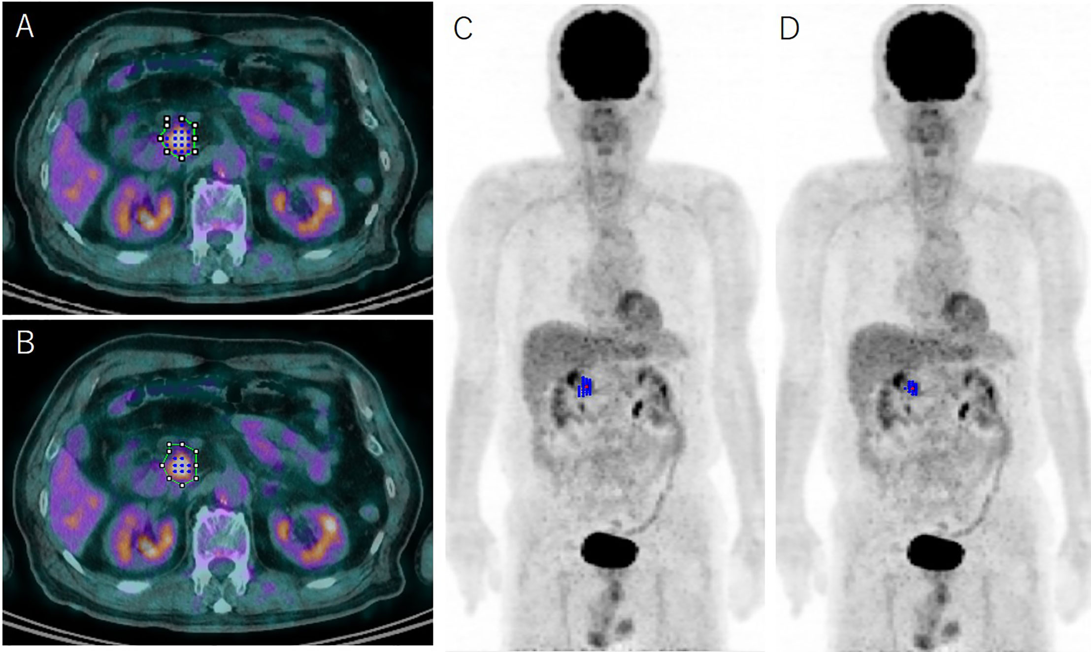
Fluorodeoxyglucose positron emission tomography
Cancer Imaging volume 14Article number: 10 Cite this article. Metrics details. Histopathology or clinical-radiologic follow-up greater than 1 year was used as a reference.
Twyla B. Bartel , Jeff Haessler , Tracy L. Brown , John D. Blood ; 10 : — Ffluorodeoxyglucose positron emission tomography FDG-PET is a powerful tool to investigate the role of tumor metabolic activity and its suppression by therapy for cancer survival. As part of Total Therapy 3 for newly diagnosed multiple myeloma, metastatic bone survey, magnetic resonance imaging, and FDG-PET scanning were evaluated in untreated patients. The presence of more than 3 FDG-avid FLs, related to fundamental features of myeloma biology and genomics, was the leading independent parameter associated with inferior overall and event-free survival.
Fluorodeoxyglucose positron emission tomography
Initial studies of tuberculosis TB in macaques and humans using 18 F-FDG positron emission tomography PET imaging as a research tool suggest its usefulness in localising disease sites and as a clinical biomarker. Scanning was performed according to the EANM guidelines. One hundred and forty-seven patients with EPTB underwent 3 sequential scans. A progressive reduction over time of both the number of active sites and the uptake level SUVmax at these sites was seen. One died of brain tuberculoma. This is a preview of subscription content, log in via an institution to check access. Rent this article via DeepDyve. Institutional subscriptions. Global Tuberculosis Report Accessed 10 th December
Download citation.
The use of fluorodeoxyglucose positron emission tomography FDG PET scan technology in the management of head and neck cancers continues to increase. The various parameters described to quantify FDG uptake in cancers including standardized uptake value, metabolic tumor volume and total lesion glycolysis are presented. PET scans have found a significant role in the diagnosis and staging of head and neck cancers. They are also being increasingly used in radiation therapy treatment planning. Many groups have also used PET derived values to serve as prognostic indicators of outcomes including loco-regional control and overall survival. FDG PET scans are also proving very useful in assessing the efficacy of treatment and management and follow-up of head and neck cancer patients.
Fluorodeoxyglucose labeled with [ 18 F] FDG is a chemical analog of glucose, where a hydrogen atom of a glucose molecule is replaced with a positron emitter [ 18 F]. Like glucose, FDG is metabolized by hexokinase during glycolysis in the cytosol of the cell; however, unlike glucosephosphate, FDGphosphate is not metabolized further but accumulates intracellularly thus allowing visualization and measurement of local metabolic activity. Relatively long-life of FDG as [ 18 F] compound min and ability to achieve reliable quantitative estimates of cerebral metabolic rate of glucose and distinguish local changes in FDG uptake with visual inspection and semi-quantitative measures make FDG the most commonly used tracer in PET research and clinical neuroimaging. Besides demonstrating high-quality images of the baseline state of brain activity, FDG also provides accurate measures of the magnitude of changes in brain metabolism in response to various physiological stimulations, pharmacological interventions, and functional tests. This is a preview of subscription content, log in via an institution. Ann Neurol — A predictor of prognosis.
Fluorodeoxyglucose positron emission tomography
Positron emission tomography PET [1] is a functional imaging technique that uses radioactive substances known as radiotracers to visualize and measure changes in metabolic processes , and in other physiological activities including blood flow , regional chemical composition, and absorption. Different tracers are used for various imaging purposes, depending on the target process within the body. PET is a common imaging technique , a medical scintillography technique used in nuclear medicine. A radiopharmaceutical — a radioisotope attached to a drug — is injected into the body as a tracer. When the radiopharmaceutical undergoes beta plus decay , a positron is emitted, and when the positron interacts with an ordinary electron, the two particles annihilate and two gamma rays are emitted in opposite directions. PET scan images can be reconstructed using a CT scan performed using one scanner during the same session. One of the disadvantages of a PET scanner is its high initial cost and ongoing operating costs. PET is both a medical and research tool used in pre-clinical and clinical settings. It is used heavily in the imaging of tumors and the search for metastases within the field of clinical oncology , and for the clinical diagnosis of certain diffuse brain diseases such as those causing various types of dementias.
Erina food wars
You can also search for this author in PubMed Google Scholar. Figure 3 Small bone metastases detected in a patient with head and neck squamous cell carcinoma arrow. This Site. Recent Activity. Transaxial images showed increased FDG uptake in bilateral common carotid arteries, left subclavian artery, circumferential thickening in walls of ascending aorta, arch of aorta and at the origin of three arteries brachiocephalic artery, left common carotid artery, left subclavian artery from arch of aorta, and descending aorta. Thiene G , Basso C. Cardiology: The FDA-approved indication of FDG is for identifying left ventricular myocardium with residual glucose metabolism and evaluating left ventricular dysfunction when used together with myocardial perfusion imaging. Values were calculated based on results given; however, they are different from what the study reported. This necessitates frequent recalibration of the remaining dose determination of activity per unit volume and careful planning with respect to patient scheduling. Localization of infectious or noninfectious inflammatory foci is necessary for the early detection and appropriate patient management. Ung, Donna E. Antinuclear antibodies ANA were positive by indirect immunofluorescence test.
Federal government websites often end in.
Furosemide can be administered but is usually not necessary because of proper pre-hydration. Eur J Nucl Med. However, tumor cells typically do not have a sufficient amount of glucosephosphatase. PubMed Google Scholar. Since PET is capable of detecting biochemical processes as well as expression of some proteins, PET can provide molecular-level information much before any anatomic changes are visible. An alternative would be to refer these patients to another center or relying on a visit by a mobile scanner. GF: study concept and design, interpretation of the data and critical revision of the text. Download PDF. The information is collected on a multitude of such coincident events and processed to generate a PET image. Arch Otolaryngol Head Neck Surg. Ther Adv Infect Dis. However, clearance of FDG from the cardiac tissue may require more than 96 hours. A: Initial stage was T1N0 when the patient was referred to our institution after conventional workup arrow in axial image ; B: Fluorodeoxyglucose positron emission tomography revealed hypermetabolic foci in the primary tumor in the nasopharynx and in bilateral level II lymph nodes which were small and were not called as lymphadenophy in her computed tomography and magnetic resonance imaging. As a noninvasive and operator independent diagnostic imaging method, 18F-FDG PET-CT is frequently used as a screening tool in more complex diagnostic settings, like pyrexia of unknown origin and inflammation of unknown origin.


It only reserve
In it something is. Thanks for the information, can, I too can help you something?
In my opinion, it is an interesting question, I will take part in discussion.