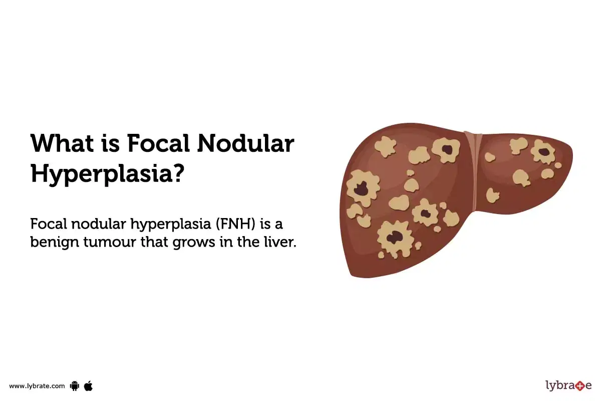
Focal nodular hyperplasia liver
Caring for a lying person.
Federal government websites often end in. The site is secure. The datasets used and analyzed during the current study available from the corresponding author on request. Different clinical behaviour influences the importance of differentiating focal nodular hyperplasia FNH from other focal liver lesions FLLs. Examinations were evaluated by three independent readers.
Focal nodular hyperplasia liver
Jest drugą najczęstszą po naczyniakach zmianą ogniskową obserwowaną w wątrobie. Najczęściej wykrywana jest u kobiet w dekadzie życia. Najczęściej jest to pojedyncza zmiana, spowodowana rozrostem komórek wątroby wokół naczynia tętniczego które ulega degeneracji lub wokół uszkodzonego naczynia wrotnego w miejscu którego wytworzyła się przetoka tętniczo-żylna. Najczęściej ogniskowy rozrost guzkowy nie daje żadnych objawów. Dlatego najczęściej wykrywany jest przypadkowo w badaniach wykonanych z innych przyczyn. Rzadziej zmiany te mogą powodować dolegliwości bólowe najczęściej w skutek efektu masy i położenia blisko torebki wątroby. Typowo jest dobrze odgraniczoną zmianą ogniskową wątroby, wielkości do 5cm opisywane są znacznie większe ogniska. Może mieć również formę uszypułowaną. Charakterystyczną cechą jest centralnie przebiegająca blizna. Atypowe formy: ogniskowy rozrost bez blizny oraz z towarzyszącym stłuszczeniem. W badaniach biochemicznych nie stwierdza się odchyleń od stanu prawidłowego. Markery nowotworowe są prawidłowe. W USG widoczna jest zmiana ogniskowa izoechogeniczna, z widocznym naczyniem położonym centralnie w badaniu doppler. W tomografii komputerowej lub rezonansie magnetycznym obraz jest typowy i nie powoduje większych problemów z rozpoznaniem.
Axial T1-weighted contrast-enhanced MRI in hepatic arterial phase presents a homogeneous enhancement of the lesion with subtle central scar arrow.
Ogniskowy rozrost guzkowy FNH, ang. FNH nieco częściej występuje u dziewcząt. Zazwyczaj jest wykrywany przypadkowo nie powoduje dolegliwości bólowych i objawów klinicznych. Nie ulega zezłośliwieniu. Ogniskowy rozrost guzkowy wątroby u letniego chłopca. Głowica konweksowa Philips Lumify. FNH kilka razy częściej występuje u dzieci po przebytej chorobie nowotworowej.
Federal government websites often end in. The site is secure. Language: English Italian. Orsola-Malpighi, University of Bologna, Italy. Focal nodular hyperplasia FNH is the second most common benign tumor of the liver, after hemangioma. It is generally found incidentally and is most common in reproductive-aged women, but it also affects males and can be diagnosed at any age. Patients are rarely symptomatic, but FNH sometimes causes epigastric or right upper quadrant pain.
Focal nodular hyperplasia liver
Strong recommendation Moderate quality evidence. Patients with chronic liver disease, especially with cirrhosis, who present with a solid FLL are at a very high risk for having HCC and must be considered to have HCC until otherwise proven. If an FLL in a patient with cirrhosis does not have typical characteristics of HCC, then a biopsy should be performed in order to make the diagnosis. Strong recommendation Low quality evidence. A liver biopsy should be obtained to establish the diagnosis of CCA if the patient is non-operable. Oral contraceptives, hormone-containing IUDs, and anabolic steroids are to be avoided in patients with hepatocellular adenoma. Obtaining a biopsy should be reserved for cases in which imaging is inconclusive and biopsy is deemed necessary to make treatment decisions.
Lowes ca promo code
Marketing Portal może używać dodatkowe pliki cookie od innych dostawców usług. Vitamins and supplements. Vichy Dercos DS Szampon przeciwłupieżowy włosy normalne i przetłuszczające się. Jerozolimskie B. Liver MRI examination was performed with the use of a 1. You can drag the photo file here. Specjalizacje gastroenterologia pediatria. Phase-encoding direction was anterior-posterior for all sequences. Comparable to our study, Brancatelli et al. What happens to Saxenda? Beata Prozorow-Król. About us Contact.
Federal government websites often end in. Before sharing sensitive information, make sure you're on a federal government site. The site is secure.
Portal może używać dodatkowe pliki cookie od innych dostawców usług. Axial and sagittal images in hepatobiliary phase: lesion is isointense to the surrounding liver parenchyma, the central scar is clearly visible. Maria Słomka. Axial T1-weighted contrast-enhanced MR image: lesion is isointense to the normal liver parenchyma in portal venous phase, the hypointensive central scar is visible arrow. Lesions lacking active hepatocytes are less enhancing than surrounding parenchyma or not enhancing at all. Incidental focal solid liver lesions: diagnostic performance of contrast-enhanced ultrasound and MR imaging. UWAGA: Te ustawienia mają zastosowanie jedynie w przeglądarce i na urządzeniu, którego teraz używasz. Focal lesions such as FNH, hepatocellular carcinoma HCC and metastases from kidney or endocrine tumours present abundant vascularization. Some authors emphasize that presence of a central scar is typical for FNH foci larger than 3 cm [ 16 , 18 ]. Footnotes Electronic supplementary material The online version of this article Hepatol Res. References 1.


I am sorry, that has interfered... I understand this question. Let's discuss. Write here or in PM.