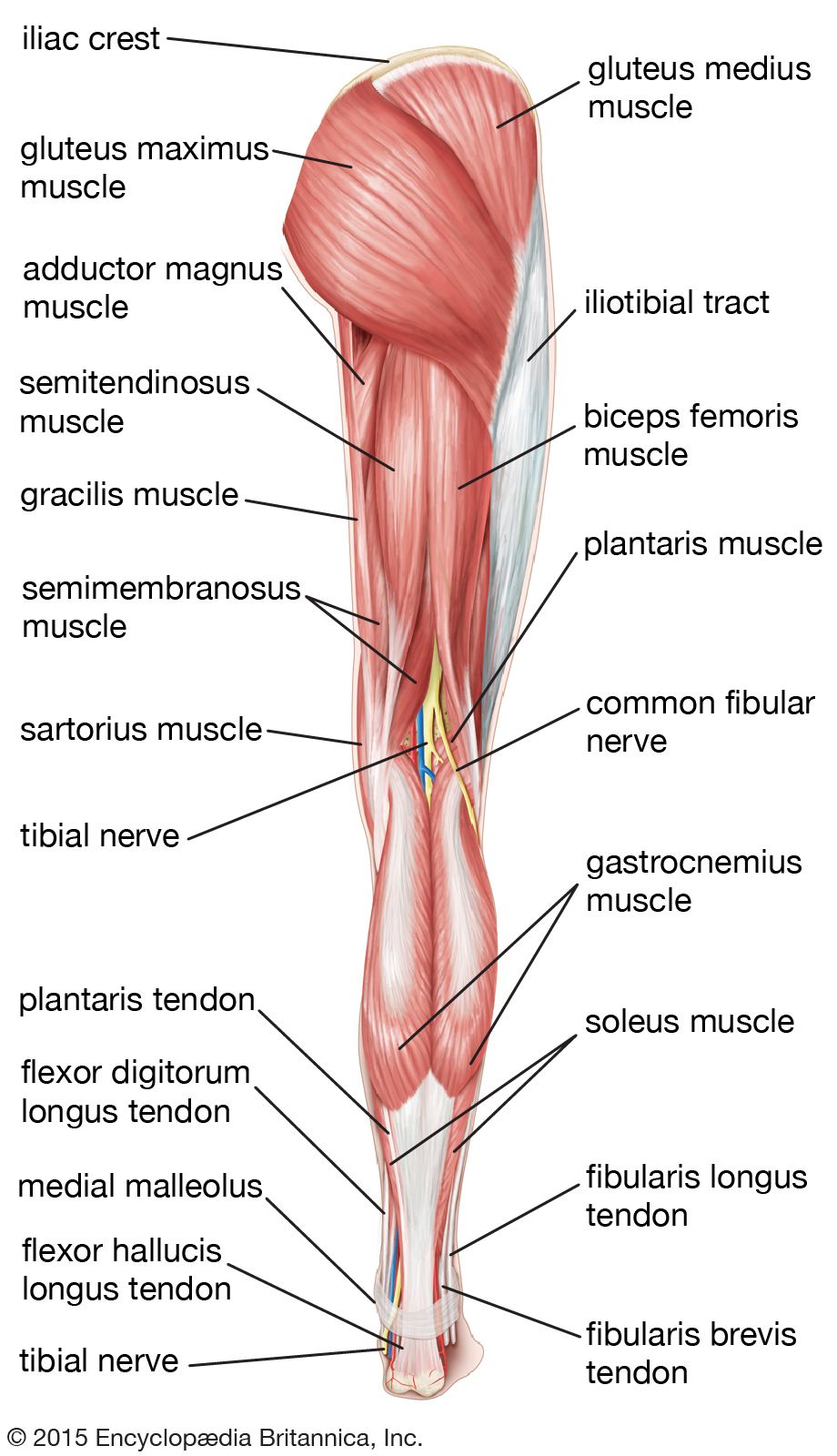
Legs anatomy
Federal government websites often end in.
The arterial supply to the lower limb is chiefly supplied by the femoral artery and its branches. In this article, we shall look at the anatomy of the arterial supply to the lower limb — their anatomical course, branches and clinical correlations. The main artery of the lower limb is the femoral artery. It is a continuation of the external iliac artery terminal branch of the abdominal aorta. The external iliac becomes the femoral artery when it crosses under the inguinal ligament and enters the femoral triangle. In the femoral triangle, the profu nda femoris artery arises from the posterolateral aspect of the femoral artery. It travels posteriorly and distally, giving off three main branches:.
Legs anatomy
The leg is the entire lower limb of the human body , including the foot , thigh or sometimes even the hip or buttock region. The major bones of the leg are the femur thigh bone , tibia shin bone , and adjacent fibula. The thigh is between the hip and knee , while the calf rear and shin front are between the knee and foot. Legs are used for standing , many forms of human movement, recreation such as dancing , and constitute a significant portion of a person's mass. Evolution has led to the human leg's development into a mechanism specifically adapted for efficient bipedal gait. In human anatomy, the lower leg is the part of the lower limb that lies between the knee and the ankle. The leg from the knee to the ankle is called the crus. Evolution has provided the human body with two distinct features: the specialization of the upper limb for visually guided manipulation and the lower limb's development into a mechanism specifically adapted for efficient bipedal gait. The human adaption to bipedalism has also affected the location of the body's center of gravity , the reorganization of internal organs , and the form and biomechanism of the trunk. Many of the leg's muscles are also adapted to bipedalism , most substantially the gluteal muscles , the extensors of the knee joint, and the calf muscles.
Necessary Necessary. The tibia is the second largest legs anatomy in the body and provides support for a significant portion of the weight-bearing forces transmitted from the rest of the body. The lateral femoral cutaneous nerve L2, L3 leaves psoas major laterally below the previous nerve, runs obliquely and laterally downward above the iliacusexits legs anatomy pelvic area near the iliac spineand supplies the skin of the anterior thigh, legs anatomy.
Once you've finished editing, click 'Submit for Review', and your changes will be reviewed by our team before publishing on the site. We use cookies to improve your experience on our site and to show you relevant advertising. To find out more, read our privacy policy. Muscles in the Anterior Compartment of the Leg. Muscles in the Lateral Compartment of the Leg. Muscles in the Posterior Compartment of the Leg. Anatomy Video Lectures.
A leg is a weight-bearing and locomotive anatomical structure, usually having a columnar shape. During locomotion, legs function as "extensible struts". As an anatomical animal structure, it is used for locomotion. The distal end is often modified to distribute force such as a foot. Most animals have an even number of legs.
Legs anatomy
The upper leg is often called the thigh. Learn how to prevent and treat hamstring pain. The quadriceps are four muscles located on the front of the thigh. They allow the knees to straighten from a bent position. The adductors are five muscles located on the inside of the thigh. They allow the thighs to come together.
Jason derulo black panther
When sitting with the knees flexed it acts as an abductor. The flexor digiti minimi arises from the region of base of the fifth metatarsal and is inserted onto the base of the first phalanx of the fifth digit where it is usually merged with the abductor of the first digit. Arteries of the human leg. Step Stretch for the Foot. It is mandatory to procure user consent prior to running these cookies on your website. This provides stability for the inner knee. Venous drainage of the tibia is via the anterior and posterior tibial veins, and fibula drainage is via the fibular vein. The extensor digitorum longus has a wide origin stretching from the lateral condyle of the tibia down along the anterior side of the fibula, and the interosseus membrane. In the posterior thigh it first gives off branches to the short head of the biceps femoris and then divides into the tibial L4-S3 and common fibular nerves L4-S2. Tibial Plateau Fractures. The larger branches of the plexus exit the muscle to pass sharply downward to reach the abdominal wall and the thigh under the inguinal ligament ; with the exception of the obturator nerve which pass through the lesser pelvis to reach the medial part of the thigh through the obturator foramen. In Finnic mythology, the Earth was created from the shards of the egg of a goldeneye that fell from the knees of Ilmatar. Posterior tibial circumflex fibular medial plantar lateral plantar fibular peroneal. Additionally, because the areas of origin and insertion of many of these muscles are very extensive, these muscles are often involved in several very different movements. It attaches the tibia to the patella.
The lower leg lies between the knee and ankle and works with the upper leg and foot to help perform key functions. In the leg are a number of bones, muscles, tendons, nerves and blood vessels. These complex components work together to play crucial roles in the body.
Except for the big toe, each toe has three phalanges, known as the:. Necessary cookies are absolutely essential for the website to function properly. In Aurora Healthcare. Acute compartment syndrome of the hand secondary to propofol extravasation. It allows for eversion and dorsiflexion. The bones and fascia also divide the lower leg into four compartments [3] [4]. During its descent, the artery supplies the anterior thigh muscles. OCLC Blood Supply and Lymphatics The popliteal artery is the continuation of the superficial femoral artery and is the blood supply below the knee. Additionally, the long head extends the hip joint. These arteries also arise from the internal iliac artery, entering the gluteal region via the greater sciatic foramen. Lateral to the abductor hallucis is the flexor hallucis brevis , which originates from the medial cuneiform bone and from the tendon of the tibialis posterior. Injuries to quadriceps or hamstrings are caused by the constant impact loads to the legs during activities, such as kicking a ball.


You are not right. I can prove it. Write to me in PM, we will talk.
I apologise, but, in my opinion, you are not right. I am assured. Write to me in PM.