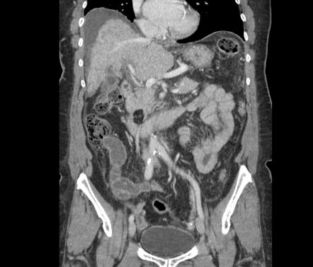
Lipoma on pancreas
Regret for the inconvenience: we are taking measures to prevent fraudulent form submissions by extractors and page crawlers.
At the time the article was last revised Daniel J Bell had no financial relationships to ineligible companies to disclose. Pancreatic lipomas are uncommon mesenchymal tumors of the pancreas. Rarely symptomatic, they are most often detected incidentally on cross-sectional imaging for another purpose. If they do cause symptoms, it will typically be those related to regional mass effect from the mass. Pancreatic lipomas are composed of mature fat cells with thin internal fibrous septa.
Lipoma on pancreas
Federal government websites often end in. The site is secure. Correspondence to: Dr. Lipomas of the pancreas are very rare. There are fewer than 25 reported cases of lipoma originating from the pancreas. We present a case of pancreatic lipoma in a year-old woman with magnetic resonance imaging findings and confirmatory histological findings. We discuss and highlight the radiological features distinguishing a pancreatic lipoma from other fatty lesions of the pancreas and pancreatic liposarcoma and provide a brief review of the literature. Pancreatic tumors arise from cells of mesenchymal or epithelial origin, or from non-ductal structures and fat. Among these, fat-originating tumors such as lipoma and liposarcomas are the rarest. A year-old woman with a past medical history of dyslipidemia and cholelithiaisis was admitted to our institution for right hypochondrium pain and epigastric discomfort and vomiting of 2 wk duration. The tumor marker carbohydrate antigen was slightly raised, Physical examination was normal. Ultrasonography revealed multiple subcentimetre gallstones; the pancreas was noted not to be well visualized due to overlying bowel gas. She was treated symptomatically for biliary colic and was discharged well with a view for an elective laparoscopic cholecystectomy.
CT findings of pancreatic lipoma: An analysis of 2 cases [in Chinese]. Rev Esp Enferm Dig ; 98 : —6. Figure 2.
Pancreatic lipomas are thought to be very rare. Lipomas are usually easy to identify on imaging, particularly via computed tomography CT. Here, we present a case of pancreatic lipoma in a year-old female. She was asymptomatic and had no medical history of note. Finally, the patient underwent a pancreaticoduodenectomy. Histologically, mature adipocytes were noted in the bulk of the tumor.
Federal government websites often end in. The site is secure. Pancreatic lipomas are rare. We present a case of incidentally discovered pancreatic lipoma in a year-old female suffering from metastatic ovarian carcinoma who was referred to radiology for follow-up imaging. Fat-containing tumours originating from the pancreas are very rare. Most lipomasshow characteristic features on imaging that allow their differentiation.
Lipoma on pancreas
Hence, localizing the tumor site can guide the healthcare provider to arrive at a probable diagnosis. The specific risk factors for Lipoma of Pancreas are unknown or unidentified. Note: It is important to note that an individual diagnosed with cancer of the pancreas may not have any of the above-mentioned risk factors. It is important to note that having a risk factor does not mean that one will get the condition. A risk factor increases ones chances of getting a condition compared to an individual without the risk factors. Some risk factors are more important than others. Also, not having a risk factor does not mean that an individual will not get the condition. It is always important to discuss the effect of risk factors with your healthcare provider. The signs and symptoms of Lipoma of Pancreas depend upon the size and location of the tumor.
Google pronounce names audio
Arch Surg. Citation, DOI, disclosures and article data. Edit article. Lipomas are most commonly found in the middle-aged and elderly, possibly because they undergo physical examinations more often than do the young. BJR Case Rep. Acta Radiol. Pediatr Med Chir. Lipomas show characteristic imaging features, identification of which allow a correct diagnosis without any histopathological confirmation. To our knowledge, our case is the first example of a suspected well-differentiated liposarcoma that was actually a pancreatic lipoma. We discuss and highlight the radiological features distinguishing a pancreatic lipoma from other fatty lesions of the pancreas and pancreatic liposarcoma and provide a brief review of the literature. Pancreatic tumors originate from mesenchymal or epithelial cells, or from non-ductal structures. Author contributions : Xiao RY collected case data, prepared the photos and wrote the manuscript; Yao X and Wang WL proofread and revised the manuscript; all of the authors approved the final version to be published. This cross sectional image reveals a 6.
Federal government websites often end in.
Butler et al. Corresponding Author of This Article. They can be found almost anywhere in the body where there is adipose tissue. Aug 26, ; 7 16 : Published online Aug 26, Rarely symptomatic, they are most often detected incidentally on cross-sectional imaging for another purpose. Detecting presence of fatty tissue, rules out the diagnosis of adenocarcinoma and pancreatic neuroendocrine tumour. Morden Medicine J China. Nonductal tumors of the pancreas. As present, imaging techniques are very accurate, and in most cases, there is no need for histopathological confirmation of pancreatic lipoma. Research Domain of This Article. BJR Case Rep. Pancreatic lipomas are uncommon mesenchymal tumors of the pancreas.


0 thoughts on “Lipoma on pancreas”