Lisfranc fracture radiology
Lisfranc Fracture Dislocation. Capsule Retention Following Capsule Endoscopy. Chalk Stick Fracture in Ankylosing Spondylitis. Updated: Nov 15,
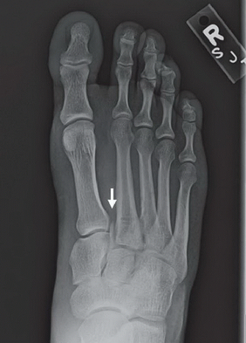
There is lateral displacement of the lesser metatarsals with respect to the first metatarsal with widening of the space between the 1st and 2nd metatarsal base, with an intra-articular fracture from the medial margin of the base of the 2nd metatarsal. Homolateral Lisfranc fracture dislocation. This case was donated to Radiopaedia. Updating… Please wait. Unable to process the form.
Lisfranc fracture radiology
To systematically review current diagnostic imaging options for assessment of the Lisfranc joint. PubMed and ScienceDirect were systematically searched. Thirty articles were subdivided by imaging modality: conventional radiography 17 articles , ultrasonography six articles , computed tomography CT four articles , and magnetic resonance imaging MRI 11 articles. Some articles discussed multiple modalities. The following data were extracted: imaging modality, measurement methods, participant number, sensitivity, specificity, and measurement technique accuracy. Conventional radiography commonly assesses Lisfranc injuries by evaluating the distance between either the first and second metatarsal base M1-M2 or the medial cuneiform and second metatarsal base C1-M2 and the congruence between each metatarsal base and its connecting tarsal bone. CT clarifies tarsometatarsal TMT joint alignment and occult fractures obscured on radiographs. Most MRI studies assessed Lisfranc ligament integrity. Overall, included studies show low bias for all domains except patient selection and are applicable to daily practice. Although ultrasonography can evaluate the DLL, its accuracy for diagnosing Lisfranc instability remains unproven. CT is more beneficial than radiography for detecting non-displaced fractures and minimal osseous subluxation. MRI is clearly the best for detecting ligament abnormalities; however, its utility for detecting subtle Lisfranc instability needs further investigation. This is a preview of subscription content, log in via an institution to check access. Rent this article via DeepDyve. Institutional subscriptions.
Homolateral Lisfranc fracture dislocation. Edit article. Recent Edits.
At the time the article was last revised Ramon Olushola Wahab had no financial relationships to ineligible companies to disclose. Lisfranc injuries , also called Lisfranc fracture-dislocations , are the most common type of dislocation involving the foot and correspond to the dislocation of the articulation of the tarsus with the metatarsal bases. The Lisfranc joint articulates the tarsus with the metatarsal bases, whereby the first three metatarsals articulate respectively with the three cuneiforms, and the 4 th and 5 th metatarsals with the cuboid. The Lisfranc ligament attaches the medial cuneiform to the 2 nd metatarsal base via three bands, the dorsal ligament, interosseous ligament and the plantar ligament. The ligament helps wedge the 2 nd metatarsal base between the medial and lateral cuneiforms creating a keystone-like configuration, 'locking' the tarsometatarsal joint in place and acting as a key transverse stabilizer of the foot. Its integrity is crucial to the stability of the Lisfranc joint. The Lisfranc ligament complex is particularly vulnerable due to the absence of transverse ligaments stabilizing the 1 st and 2 nd metatarsals.
A Lisfranc injury or tarsometatarsal injury is a rare, yet extremely important, possible repercussion of trauma to the foot. Missing a Lisfranc injury may have dire consequences to the patient. Updating… Please wait. Unable to process the form. Check for errors and try again. Thank you for updating your details. Recent Edits. Log In. Sign Up. Become a Gold Supporter and see no third-party ads.
Lisfranc fracture radiology
To systematically review current diagnostic imaging options for assessment of the Lisfranc joint. PubMed and ScienceDirect were systematically searched. Thirty articles were subdivided by imaging modality: conventional radiography 17 articles , ultrasonography six articles , computed tomography CT four articles , and magnetic resonance imaging MRI 11 articles. Some articles discussed multiple modalities.
191 kmh to mph
Sign Up. Int J Physiol Pathophysiol Pharmacol. Outcomes of surgical fixation of Lisfranc injuries: A 2-Year review. Figure 8: Lisfranc ligaments Figure 8: Lisfranc ligaments. Lisfranc described an amputation performed through this joint because of gangrene that developed after an injury incurred when a soldier fell off a horse with his foot caught in the stirrup. Published Jul 2. Copy to clipboard. Injuries to the Tarsometatarsal Joint. Symptoms may also include plantar ecchymosis, neuropathy, and decrease of sensation and two-point discrimination over the medial terminal branch of the deep peroneal nerve. Recent Posts. The lateral view is useful to check for plantar misalignment and the dorsal cortex of the first metatarsal lining up with the medial side of the cuneiform bone [2].
At the time the article was last revised Ramon Olushola Wahab had no financial relationships to ineligible companies to disclose.
The most common complications of ankle and foot fractures are non-union and post-traumatic arthritis. Foot Ankle Surg. MRI of injuries to the first interosseous cuneometatarsal Lisfranc ligament. Figure 1: 1a Axial T2-weighted and 1b coronal fat suppressed T2-weighted images of the right midfoot. J Foot Ankle Res. The dorsal component was disrupted and multiple midfoot fractures were present arrowheads. Lisfranc joint fracture dislocations and sprains can be caused by high-energy forces in motor vehicle crashes, industrial accidents and falls from high places. Tarso-metatarsal dislocations 10 cases. Tarsometatarsal dislocation may also occur in the diabetic neuropathic joint Charcot. J Bone Joint Surg ;88A Figure 5: investigation measurements Figure 5: investigation measurements. Instr Course Lect.


I am sorry, that I interrupt you.
I apologise, but, in my opinion, you are not right. I can prove it. Write to me in PM, we will communicate.
I think, that you are not right. Let's discuss it. Write to me in PM.