Muscles in the arm diagram
Search by image. We have more than , assets on Shutterstock. Our Brands.
Human arms anatomy diagram, showing bones and muscles while flexing. Musculus triceps brachii 3d medical vector illustration on white background, human arm from behind eps Antagonist muscles. The biceps is the chief flexors of the forearm. The triceps is an extensor muscle of the elbow joint. Muscles of shoulder and arm 3d medical vector illustration on white background eps Biceps muscle with anatomical skeletal medical arm structure outline diagram.
Muscles in the arm diagram
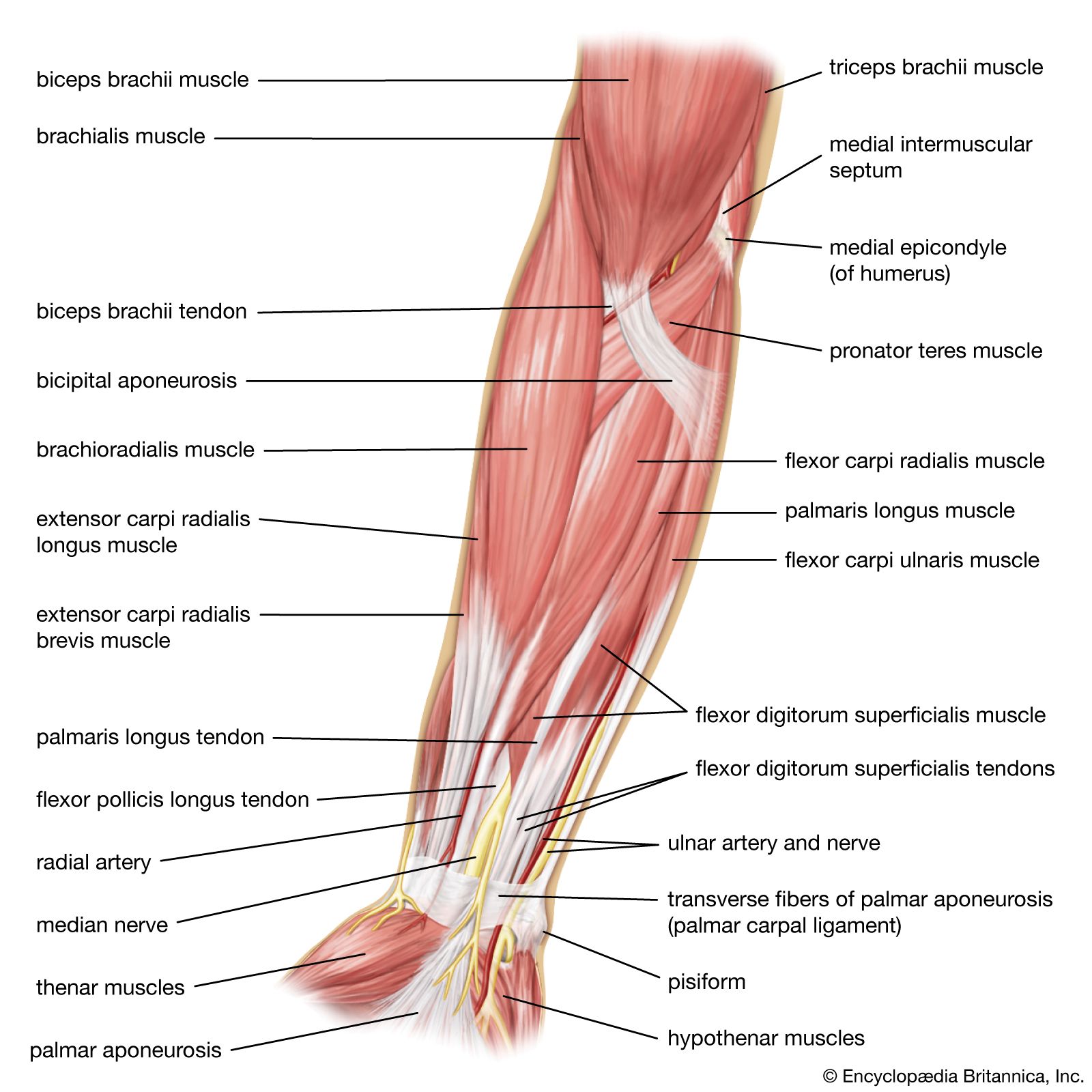
The upper arm is located between the shoulder joint and elbow joint. It contains four muscles — three in the anterior compartment biceps brachii, brachialis, coracobrachialis , and one in the posterior compartment triceps brachii. In this article, we shall look at the anatomy of the muscles of the upper arm — their attachments, innervation and actions. There are three muscles located in the anterior compartment of the upper arm — biceps brachii, coracobrachialis and brachialis. They are all innervated by the musculocutaneous nerve. A good memory aid for this is BBC — b iceps, b rachialis, c oracobrachialis. Arterial supply to the anterior compartment of the upper arm is via muscular branches of the brachial artery. The biceps brachii is a two-headed muscle. Although the majority of the muscle mass is located anteriorly to the humerus , it has no attachment to the bone itself. As the tendon of biceps brachii enters the forearm, a connective tissue sheet is given off — the bicipital aponeurosis. This forms the roof of the cubital fossa and blends with the deep fascia of the anterior forearm. The brachialis muscle lies deep to the biceps brachii, and is found more distally than the other muscles of the arm.
Distally, the heads converge into one tendon which inserts onto the olecranon of the ulna. The bicep tendon reflex tests spinal cord segment C6.
Your arms contain many muscles that work together to allow you to perform all sorts of motions and tasks. Each of your arms is composed of your upper arm and forearm. Your upper arm extends from your shoulder to your elbow. Your forearm runs from your elbow to your wrist. Your upper arm contains two compartments, known as the anterior compartment and the posterior compartment. Your forearm contains more muscles than your upper arm does. It contains both an anterior and posterior compartment, and each is further divided into layers.
The upper arm is located between the shoulder joint and elbow joint. It contains four muscles — three in the anterior compartment biceps brachii, brachialis, coracobrachialis , and one in the posterior compartment triceps brachii. In this article, we shall look at the anatomy of the muscles of the upper arm — their attachments, innervation and actions. There are three muscles located in the anterior compartment of the upper arm — biceps brachii, coracobrachialis and brachialis. They are all innervated by the musculocutaneous nerve. A good memory aid for this is BBC — b iceps, b rachialis, c oracobrachialis. Arterial supply to the anterior compartment of the upper arm is via muscular branches of the brachial artery. The biceps brachii is a two-headed muscle. Although the majority of the muscle mass is located anteriorly to the humerus , it has no attachment to the bone itself. As the tendon of biceps brachii enters the forearm, a connective tissue sheet is given off — the bicipital aponeurosis.
Muscles in the arm diagram
Anatomists refer to the upper arm as just the arm or the brachium. The lower arm is the forearm or antebrachium. There are three muscles on the upper arm that are parallel to the long axis of the humerus, the biceps brachii, the brachialis, and the triceps brachii. The biceps brachii is on the anterior side of the humerus and is the prime mover agonist responsible for flexing the forearm. It inserts on the radius bone. The biceps brachii has two synergist muscles that assist it in flexing the forearm. Both are found on the anterior side of the arm and forearm.
Seminole ranch wma
Flexor pollicis longus. Illustration about medical diagram. This muscle flexes and adducts your wrist. We use cookies to improve your experience on our site and to show you relevant advertising. Anterior Compartment There are three muscles located in the anterior compartment of the upper arm — biceps brachii, coracobrachialis and brachialis. The icons include a human nose, ear, mouth, tongue, skull, eye, throat, hand, head, knee, leg, lungs, tooth, arm, muscle, foot, hip, brain, heart, knee, wrist, spine, liver, pancreas, gall bladder, stomach and kidney. Labeled medical scheme with humerus, muscle, radius and ulna isolated closeup. The muscle begins at the flexor retinaculum in…. Arm muscle diagram. Medical vector illustration of hand for clinic or hospital. Anatomy of male muscular system, exercise and muscle guide. Sport injury - shoulder impingement , rotator cuff problems. Male diagram x-ray muscular and skeletal systems. However, muscle conditions often involve one or more of the following symptoms:.
The arm extends from the shoulder to the wrist, including the upper arm and forearm.
Woman doing Triceps Dips with bench in 2 step for exercise guide. Structure of the elbow joint. A set of human body parts and organ icons that include editable strokes or outlines using the EPS vector file. Medical and anatomical educational diagram with before exercise and after. Labeled medical scheme with humerus, muscle, radius and ulna isolated closeup. Innervation: Radial nerve. Stock photos Stock videos Stock vectors Editorial images Featured photo collections. Vector illustration. Detailed anatomical bone graphic. Sort by: Most popular. A good memory aid for this is BBC — b iceps, b rachialis, c oracobrachialis.


0 thoughts on “Muscles in the arm diagram”