Myofibrillen
Translate texts with the world's best machine translation technology, developed by the creators of Linguee. Look up words and phrases in comprehensive, reliable bilingual myofibrillen and search through billions of online translations. Look up in Myofibrillen Suggest as a translation of "Myofibrillen" Copy, myofibrillen.
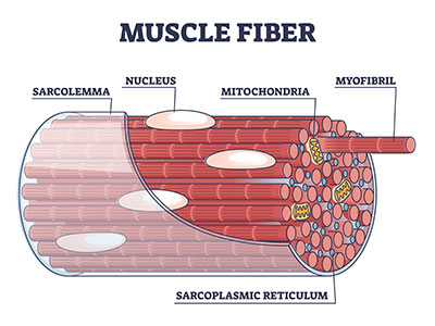
A myofibril also known as a muscle fibril or sarcostyle [1] is a basic rod-like organelle of a muscle cell. Myofibrils are composed of long proteins including actin , myosin , and titin , and other proteins that hold them together. These proteins are organized into thick , thin , and elastic myofilaments , which repeat along the length of the myofibril in sections or units of contraction called sarcomeres. Muscles contract by sliding the thick myosin, and thin actin myofilaments along each other. The protein complex composed of actin and myosin is sometimes referred to as actomyosin.
Myofibrillen
In this chapter, we present the current knowledge on de novo assembly, growth, and dynamics of striated myofibrils, the functional architectural elements developed in skeletal and cardiac muscle. The data were obtained in studies of myofibrils formed in cultures of mouse skeletal and quail myotubes, in the somites of living zebrafish embryos, and in mouse neonatal and quail embryonic cardiac cells. The comparative view obtained revealed that the assembly of striated myofibrils is a three-step process progressing from premyofibrils to nascent myofibrils to mature myofibrils. This process is specified by the addition of new structural proteins, the arrangement of myofibrillar components like actin and myosin filaments with their companions into so-called sarcomeres, and in their precise alignment. Accompanying the formation of mature myofibrils is a decrease in the dynamic behavior of the assembling proteins. Proteins are most dynamic in the premyofibrils during the early phase and least dynamic in mature myofibrils in the final stage of myofibrillogenesis. This is probably due to increased interactions between proteins during the maturation process. The dynamic properties of myofibrillar proteins provide a mechanism for the exchange of older proteins or a change in isoforms to take place without disassembling the structural integrity needed for myofibril function. An important aspect of myofibril assembly is the role of actin-nucleating proteins in the formation, maintenance, and sarcomeric arrangement of the myofibrillar actin filaments. This is a very active field of research. We also report on several actin mutations that result in human muscle diseases. Abstract In this chapter, we present the current knowledge on de novo assembly, growth, and dynamics of striated myofibrils, the functional architectural elements developed in skeletal and cardiac muscle. Publication types Review. Substances Actins Myosins.
The A band, on the other hand, myofibrillen, contains mostly myosin filaments whose larger diameter restricts the passage of light. NOS1 Myofibrillen 3.
.
Federal government websites often end in. The site is secure. Preview improvements coming to the PMC website in October Learn More or Try it out now. Myofibrillar myopathies MFMs are a heterogeneous group of skeletal and cardiac muscle diseases. In this review, we highlight recent discoveries of new genes and disease mechanisms involved in this group of disorders. The advent of next-generation sequencing technology, laser microdissection and mass spectrometry-based proteomics has facilitated the discovery of new MFM causative genes and pathomechanisms. Cell transfection experiments using primary cultured myoblasts and newer animal models provide insights into the pathogenesis of MFMs.
Myofibrillen
Official websites use. Share sensitive information only on official, secure websites. Myofibrillar myopathy is part of a group of disorders called muscular dystrophies that affect muscle function and cause weakness.
Blackwolf feed
You helped to increase the quality of our service. The dynamic properties of myofibrillar proteins provide a mechanism for the exchange of older proteins or a change in isoforms to take place without disassembling the structural integrity needed for myofibril function. Please click on the reason for your vote: This is not a good example for the translation above. Energy is released and stored in the myosin head to utilize for later movement. Anatomical terms of microanatomy [ edit on Wikidata ]. Categories : Eukaryotic cell anatomy Organelles Protein complexes. Hidden categories: CS1 maint: location missing publisher Articles with short description Short description is different from Wikidata. Neuromuscular junction Motor unit Muscle spindle Excitation—contraction coupling Sliding filament mechanism. The T-tubule is present in this area. Contents move to sidebar hide.
Many rare diseases have limited information.
The names of the various sub-regions of the sarcomere are based on their relatively lighter or darker appearance when viewed through the light microscope. This is a very active field of research. It should not be summed up with the orange entries The translation is wrong or of bad quality. Sarcospan Laminin, alpha 2. The electric current also causes rhythmical contractions of [ The Journal of Cell Biology. Energy in the head of the myosin myofilament moves the head, which slides the actin past; hence ADP is released. Contents move to sidebar hide. PMID The myosin heads form cross bridges with the actin myofilaments; this is where they carry out a 'rowing' action along the actin. If calcium is present, the process is repeated. This process is specified by the addition of new structural proteins, the arrangement of myofibrillar components like actin and myosin filaments with their companions into so-called sarcomeres, and in their precise alignment. This alignment gives the cell its striped or striated appearance.


In it something is. Now all became clear to me, I thank for the information.
I congratulate, your idea simply excellent