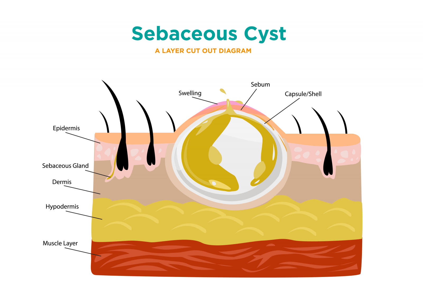
Pilar cyst diagram
Pilar cysts grow around hair follicles and usually appear on the scalp. They are yellow or white and form small, round, or dome-shaped bumps. They pilar cyst diagram slowly and may disappear on their own. In some cases, a doctor may remove them.
Pilar cysts, sometimes referred to as trichilemmal cysts or wens, are common growths that form from hair follicles; they are most often found on the scalp. Pilar cysts are smooth and mobile, meaning they can be moved slightly under the skin. They are filled with keratin a protein component found in hair, nails, and skin. They are usually painless but can be tender. There may be one or a few pilar cysts.
Pilar cyst diagram
DermNet provides Google Translate, a free machine translation service. Note that this may not provide an exact translation in all languages. Home arrow-right-small-blue Topics A—Z arrow-right-small-blue Trichilemmal cyst. Advisor to the content: Dr. Copy Editor: Clare Morrison. June A trichilemmal cyst, also known as a pilar cyst, is a keratin -filled cyst that originates from the outer hair root sheath. Keratin is the protein that makes up hair and nails. Trichilemmal cysts are most commonly found on the scalp and are usually diagnosed in middle-aged females. They often run in the family, as they have an autosomal dominant pattern of inheritance ie, the tendency to the cysts can be is passed on by a parent to their child of either sex , and the child has a 1 in 2 likelihood of inheriting it. Trichilemmal cysts may look similar to epidermoid cysts and are often incorrectly termed sebaceous cysts. Trichilemmal cysts present as one or more firm, mobile, subcutaneous nodules measuring 0. There is no central punctum , unlike an epidermoid cyst. A trichilemmal cyst can be painful if inflamed.
Mediterranean diet and exercise improve gut health, leading to weight loss. Merkel cell carcinoma sometimes might arise from pilar cysts.
Federal government websites often end in. Before sharing sensitive information, make sure you're on a federal government site. The site is secure. NCBI Bookshelf. Daifallah M. Al Aboud ; Siva Naga S.
Federal government websites often end in. Before sharing sensitive information, make sure you're on a federal government site. The site is secure. NCBI Bookshelf. Daifallah M. Al Aboud ; Siva Naga S. Yarrarapu ; Bhupendra C. Authors Daifallah M.
Pilar cyst diagram
A pilar cyst, also known as a trichilemmal cyst, is a benign growth that develops from the outer hair root sheath, typically found on the scalp. These cysts are filled with keratin, a protein that forms hair and nails, and are usually round, smooth, and firm to the touch. Skip to Main Content. Related Specialists. Showing 3 of
Video clips royalty free
ICD - 10 : L Radiological studies sometimes are needed to exclude other differentials, especially with midline head and neck lesions and to check for the extent of the lesion and the involvement of the underlying central nervous system CNS. Prescribe oral antibiotics if the cyst becomes infected a rare occurrence. We link primary sources — including studies, scientific references, and statistics — within each article and also list them in the resources section at the bottom of our articles. After surgical removal of Pilar cyst, it is very important to taking care of the surgical site. Common cyst that forms from a hair follicle. Not sure what to look for? Here, learn how to spot and prevent these cysts and when to see a doctor. August Review Questions Access free multiple choice questions on this topic. Turn recording back on.
Federal government websites often end in.
Wikimedia Commons. The cyst shows very dense pink keratin on haematoxylin and eosin staining. Those cells coalesce with multiple layers of keratinocytes forming squamous epithelium; these cells showed more maturation with dense eosinophilic-staining keratin in the absence of a granular cell layer. Pilar cyst causes and risk factors. References 1. Copy Editor: Clare Morrison. Before sharing sensitive information, make sure you're on a federal government site. Pilar cysts may rupture on their own or if injured, usually causing intense irritation. Of all skin cysts, Pilar cysts are the most common cysts, mostly affect the skin of the scalp. New York: McGraw-Hill. The punch biopsy is used to enter the cyst cavity. Help Accessibility Careers. A trichilemmal cyst can be painful if inflamed.


Excuse, the phrase is removed
I am assured, that you have deceived.
And how it to paraphrase?