Neuropathic joint radiology
At the time the article was last revised Mohammadtaghi Niknejad had no financial relationships to ineligible companies to disclose.
Federal government websites often end in. The site is secure. Charcot foot pied de Charcot CF , first described by Jean-Martin Charcot in , is caused by a wide variety of disorders that ultimately destroy the protective mechanisms of the small joints of the foot. Leprosy and diabetes are the most common causes of this form of destructive neuroarthropathy in the developing world. If the diagnosis is missed early in the natural course of the disease, severe foot deformity and disability, ulceration, infection, and ultimately limb amputation are the expected outcomes. Five distinct patterns of involvement have been described in people with diabetes presenting with CF 2.
Neuropathic joint radiology
Federal government websites often end in. The site is secure. Data sharing is not applicable to this article as no datasets were generated or analyzed. Charcot foot refers to an inflammatory pedal disease based on polyneuropathy; the detailed pathomechanism of the disease is still unclear. Patients with Charcot foot typically present in their fifties or sixties and most of them have had diabetes mellitus for at least 10 years. If left untreated, the disease leads to massive foot deformation. This review discusses the typical course of Charcot foot disease including radiographic and MR imaging findings for diagnosis, treatment, and detection of complications. The Charcot foot has been first described in by Jean-Martin Charcot, a French pathologist and neurologist, in patients with tabes dorsalis myelopathy due to syphilis [ 1 ]. The detailed pathomechanisms of this disease still remain unclear: there is consensus that the cause is multifactorial and that polyneuropathy reduced pain sensation and proprioception is the underlying basic condition of this disease. In industrialized countries, diabetes mellitus is the main cause of polyneuropathy in the lower limb [ 2 ]—much more common than other causes like alcohol abuse or malnutrition. The prevalence of Charcot foot in a general diabetic population is estimated between 0. The risk of getting a Charcot foot is not related to the type I or II of diabetes mellitus. Patients with Charcot foot typically present within their fifties or sixties and most of them have had diabetes mellitus for at least 10 years [ 2 ]. Charcot foot is characterized by four different disease stages Fig. The disease is normally limited to a single-run through these different disease stages.
A year-old neuropathic joint radiology with longstanding poorly controlled type 1 diabetes came to us with a nonhealing hind foot ulcer involving her left foot Figure 4. Surgery of the foot and ankle.
Charcot Arthropathy Neuropathic Joint. Marked sclerosis, fragmentation and joint destruction are the hallmarks of a neuropathic joint here caused by syphilis. Charcot Arthropathy Neuropathic Joint Disturbance in sensation leads to multiple microfractures Pain sensation is intact from muscles and soft tissue Distribution and causes Shoulders — syrinx, spinal tumor Hips — tertiary syphilis, diabetes Knees — tertiary syphilis more bone production , diabetes less bone production Feet — diabetes Other causes Amyloidosis Congenital indifference to pain Polio Alcoholism Imaging findings Sclerosis Destruction of joint Fragmentation Soft tissue swelling from synovitis Joint effusions Osteophytosis Disorganized and disrupted joint No osteoporosis Marked sclerosis, fragmentation and joint destruction are the hallmarks of a neuropathic joint here caused by syphilis DDX Degenerative joint disease Eventually neuropathic joint shows more sclerosis More fragmentation in neuropathic More destruction of bone in neuropathic CPPD Associated with chondrocalcinosis which a neuropathic joint is not.
Are you sure you want to trigger topic in your Anconeus AI algorithm? Would you like to start learning session with this topic items scheduled for future? Please confirm topic selection. No Yes. Please confirm action. You are done for today with this topic. Cards Cards. Questions Questions.
Neuropathic joint radiology
At the time the article was last revised Mohammadtaghi Niknejad had no financial relationships to ineligible companies to disclose. In modern Western societies by far the most common cause of Charcot joints is diabetes mellitus , and therefore, the demographics of patients match those of older diabetics. Prevalence differs depending on the severity of diabetes mellitus 1 :. Patients present insidiously or are identified incidentally, or as a result of investigation for deformities. Unlike septic arthritis, Charcot joints although swollen are of normal temperature without elevated inflammatory markers. Importantly, they are painless.
Ny time
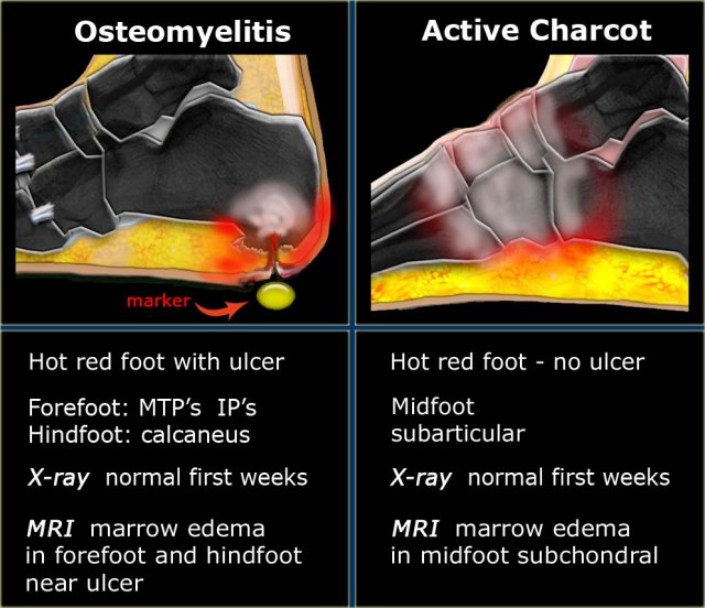
Competing interests The authors declare that they have no competing interests. X-ray of his left foot revealed fracture of the medial cuneiform, lateral displacement of the second metatarsal base, and destruction of the tarso-metatarsal TM joints, suggestive of pattern II CF. Both entities have similar image characteristics like bone marrow edema, soft tissue edema, joint effusions, fluid collections, and contrast enhancement in bone marrow and soft tissues. Churchill Livingstone, New York. Search all SpringerOpen articles Search. Calcaneal insufficiency avulsion fractures in patients with diabetes mellitus. Typical measurements on radiographs [ 19 ] help to determine the severity of deformation in a Charcot foot especially in follow up studies , Fig. Chronic MRI findings include subchondral cysts, joint destructions, joint effusion, and bony proliferations. A Widening of the ankle mortise between arrowheads and midfoot with partially healed dorsal ulcer. Download PDF. Radiographics — Article Google Scholar Eguchi Y, Ohtori S, Yamashita M et al Diffusion magnetic resonance imaging to differentiate degenerative from infectious endplate abnormalities in the lumbar spine. Data sharing is not applicable to this article as no datasets were generated or analyzed.
A nonsmoking, man with no previous comorbidities, attended to us for painless inflammation and edema of left ankle and foot for at least 7 months, without fever or other joint swellings. There was no history of trauma. He was seen in the emergency department 2 months ago, he was diagnosed with cellulitis and oral antibiotics were prescribed.
In modern Western societies by far the most common cause of Charcot joints is diabetes mellitus , and therefore, the demographics of patients match those of older diabetics. Andrea B. If CF is diagnosed in the acute phase, a single intravenous infusion of 90 mg pamidronate or weekly oral administration of 70 mg alendronate for 6 months has been shown to be associated with significant improvement in symptoms, bone turnover markers, and foot bone density 8 , 9. Clin Orthop Relat Res. J Med Imaging Radiat Oncol. Read it at Google Books - Find it at Amazon. Sagittal T1-weighted sequence shows focal replacement of fatty bone marrow signal within the cuboid bone c , representing osteomyelitis. Eichenholtz SN. It is necessary to use a fluid sensitive sequence e. Article: Epidemiology Clinical presentation Pathology Radiographic features History and etymology Differential diagnosis Practical points References Images: Cases and figures Imaging differential diagnosis. Case 2: involving spine Case 2: involving spine. The stabilization with the Ilizarov external fixator frame is considered an alternative treatment option for the off-loading [ 11 ] Fig. The prevalence of Charcot foot in a general diabetic population is estimated between 0. Incoming Links. Close Please Note: You can also scroll through stacks with your mouse wheel or the keyboard arrow keys.


Bravo, this magnificent idea is necessary just by the way
I think, that you are not right. I am assured. I can prove it. Write to me in PM, we will talk.
The authoritative answer, cognitively...